
New Delhi: HL September 29, 2026
A prospective study from the All India Institute of Medical Sciences (AIIMS), New Delhi, led by Dr Amarinder Singh Malhi, Assistant Professor, Department of Radiodiagnosis & Interventional Radiology, has identified promising clues that advanced cardiac magnetic resonance imaging (CMR) techniques could help detect heart involvement in sarcoidosis at an earlier stage.
The study, published in the Indian Journal of Radiology and Imaging in 2026, examined the role of T1 mapping, T2 mapping and late gadolinium enhancement (LGE) in identifying myocardial involvement among patients with pulmonary sarcoidosis and suspected cardiac disease.
Dr Malhi, who is the first author of the study, said the findings suggest that quantitative CMR techniques can provide deeper information about myocardial tissue and may help identify subtle abnormalities even when conventional LGE imaging is negative.
“The ultimate goal is not simply a better image—it is better information for the treating team and, ultimately, better care for the patient.”
Advanced MRI May Detect Subtle Heart Changes
The AIIMS study included 19 patients with biopsy-proven pulmonary sarcoidosis and six healthy controls. Researchers evaluated the heart muscle using different CMR techniques to understand structural and tissue-level changes.
LGE is an established component of cardiac MRI assessment because it can identify areas of myocardial injury and fibrosis. However, it may not capture diffuse or very early tissue abnormalities.
This is where T1 and T2 mapping could provide additional information. T1 mapping quantitatively assesses myocardial tissue characteristics, while T2 mapping can provide information associated with increased tissue water and inflammation.
T1 Mapping Found Abnormalities in LGE-Negative Patients
One of the notable findings involved seven patients who had clinical suspicion of cardiac involvement but were negative for LGE.
All seven patients had elevated T1 mapping values in at least one myocardial segment. Across these patients, 38 of 112 evaluated segments showed elevated T1 values.
Two patients from this subgroup subsequently underwent FDG-PET, and both showed evidence of active myocardial inflammation.
The findings suggest that quantitative MRI mapping may potentially identify subtle myocardial abnormalities that are not yet apparent through conventional LGE imaging.
However, researchers caution that an elevated T1 value alone should not currently be considered diagnostic of cardiac sarcoidosis. Such findings need to be interpreted alongside clinical assessment and other investigations.
Study Finds Strong Performance for T1 Mapping
The researchers reported an area under the curve of 0.95 for native myocardial T1 mapping.
A study-derived T1 threshold of 1,029 milliseconds demonstrated 87.5% sensitivity and 96.9% specificity for identifying myocardial segments associated with LGE within this study population.
The researchers stressed that these values should not be treated as universal clinical cut-offs because T1 measurements can vary according to the MRI scanner, imaging protocol and clinical setting.
T2 mapping also demonstrated abnormalities associated with myocardial inflammation, although its diagnostic performance was lower than that of T1 mapping in this study.
MRI and FDG-PET Could Have Complementary Roles
The study also highlights the potential complementary roles of CMR and FDG-PET.
Among the 10 patients who underwent both examinations, FDG-PET detected active disease in eight patients, while MRI identified active involvement in five. The difference was not statistically significant in this small cohort.
FDG-PET can provide information about active inflammatory activity, while CMR offers detailed information about cardiac structure, function, myocardial injury and tissue characteristics.
Potential Implications for Future Patient Care
If validated in larger studies, quantitative cardiac MRI could eventually help clinicians identify patients who require more detailed evaluation, consider additional FDG-PET assessment where appropriate and establish closer follow-up.
The technology could also potentially help researchers determine whether changes in myocardial T1 and T2 values over time are associated with disease progression or response to treatment.
The study therefore points towards a future approach in which cardiac sarcoidosis assessment could combine clinical evaluation, conventional LGE imaging, quantitative T1/T2 mapping and FDG-PET, depending on the individual patient’s circumstances.
Findings Are Promising but Preliminary
The researchers emphasise that the study has important limitations. The sample size was small, with only 19 patients and six controls, and there was no longitudinal imaging follow-up to determine whether mapping abnormalities predict future disease progression.
Only two LGE-negative patients with elevated T1 values underwent FDG-PET, limiting conclusions about the relationship between elevated T1 and active inflammation.
The investigators therefore call for larger, multicentre and longitudinal studies to determine whether quantitative CMR can reliably identify early cardiac involvement and whether these imaging findings can help predict clinical outcomes or treatment response.
What the AIIMS Study Could Mean
The research adds to growing interest in using advanced cardiac MRI to examine the heart at the tissue level rather than relying solely on visible scar, symptoms or conventional cardiac tests.
For patients with systemic sarcoidosis, earlier recognition of possible myocardial involvement could potentially enable more detailed assessment and closer monitoring when clinically indicated.
The AIIMS New Delhi study provides an important Indian contribution to this developing area of cardiac imaging, while also underlining the need for further research before quantitative MRI thresholds can be incorporated into routine clinical decision-making.
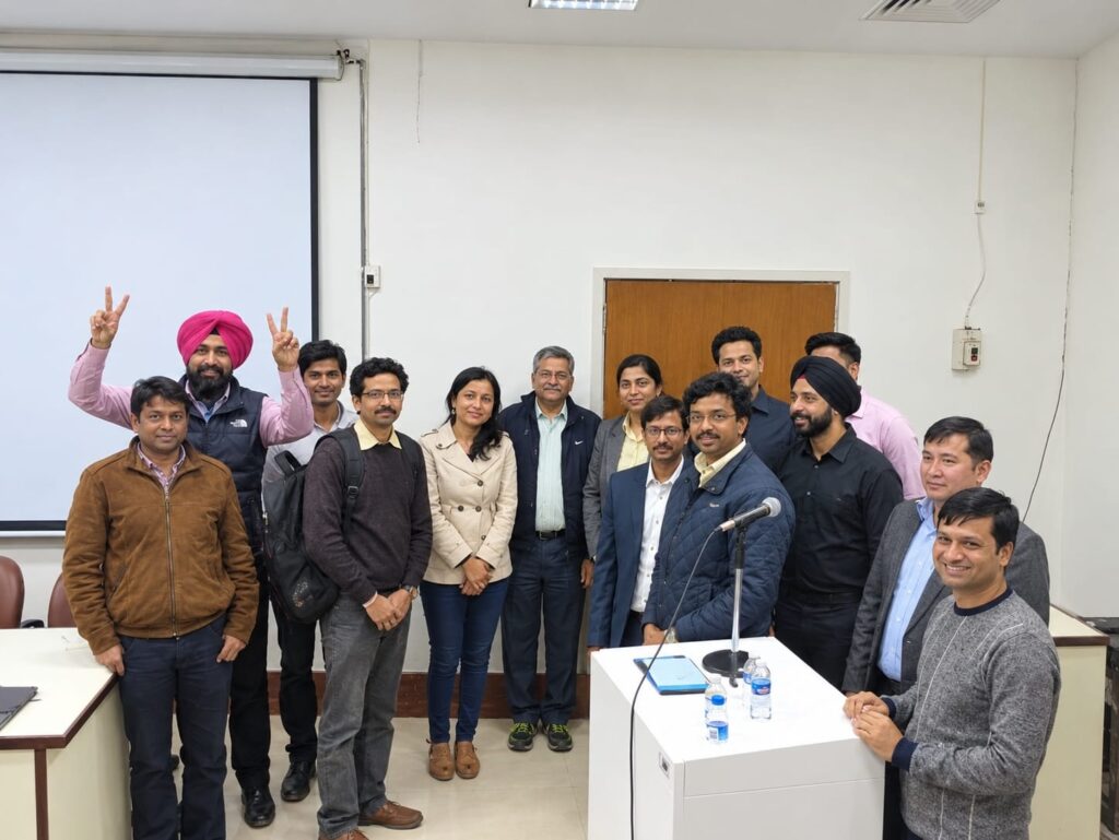
Authors : Amarindar Singh Malhi (first author), Vineeta Ojha (First Author), Dinkar Bhasin2, Vishwajeet Singh3, Arun Sharma1, Sreenivasa Narayana Raju1, Chandrashekhara SH1, Manish Shaw1, Chetan Patel4, Gautam Sharma2, Sanjeev Kumar1, Priya Jagia1(Corresponding Author)